Tools for efficient analysis of neurons in a 3D reference atlas of whole mouse spinal cord
Felix Fiederling, Luke A. Hammond, David Ng, Carol Mason & Jane Dodd (2021) Cell Reports Methods
To fill the prevailing gap in methodology for whole spinal cord analysis, we have developed a new histological and image analysis pipeline that includes:
SpineRacks - scaffolds that facilitate efficient and ordered cryo-sectioning of the entire SC in a single block
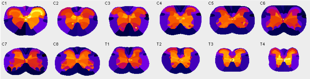
A 3D reference atlas of adult mouse SC
SpinalJ - a software tool based in Fiji to register images of sections and for standardized analysis of cells and projections in atlas space
We have verified mapping accuracies for known neurons and demonstrated the usefulness of this platform to reveal unknown neuronal distributions. Together, these tools provide high-throughput analyses of whole mouse SC and enable direct comparison of 3D spatial information between animals and studies.
This is a collaboration between Cellular Imaging and the labs of Jane Dodd and Carol Mason.
This project used the AZ100 slide scanner for imaging, with automation provided by NIS-Elements JOBS and General Analysis. It builds upon Cellular Imaging’s BrainJ project.